
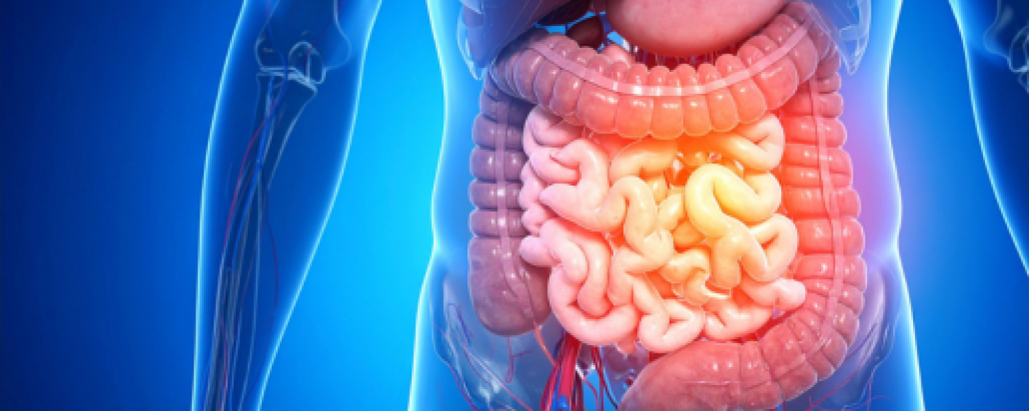
If you’re struggling with Long COVID, POTS, ME/CFS, or any lingering post-viral illness, you’ve probably felt like your body has been hijacked due to extreme exhaustion, brain fog, and strangely “off” in ways doctors sometimes struggle to explain.
Could a hidden piece of the puzzle be something as fundamental as serotonin? Not the serotonin in your brain, but the vast, separate pool outside the CNS produced in your gut.Peripheral serotonin plays critical roles in energy production, blood flow, vagal nerve activation, gut function, control of inflammation, and even mitochondrial health.
In this article, we will review emerging research showing that circulating serotonin is dangerously depleted in many people with these exact conditions.
Serotonin Synthesis Beyond the Brain
Serotonin outside the central nervous system (CNS) is primarily synthesized in the gastrointestinal tract by enterochromaffin cells in the intestinal mucosa. These peripheral sources account for roughly 90% of the body’s total serotonin. However, the serotonin made in the gut can’t cross the blood-brain barrier to enter the CNS. Thus, peripheral serotonin represents a second, much larger pool in the body that plays an important role in our physiology but is unrelated to its function as a neurotransmitter in the brain.
Serotonin Biosynthesis From Tryptophan
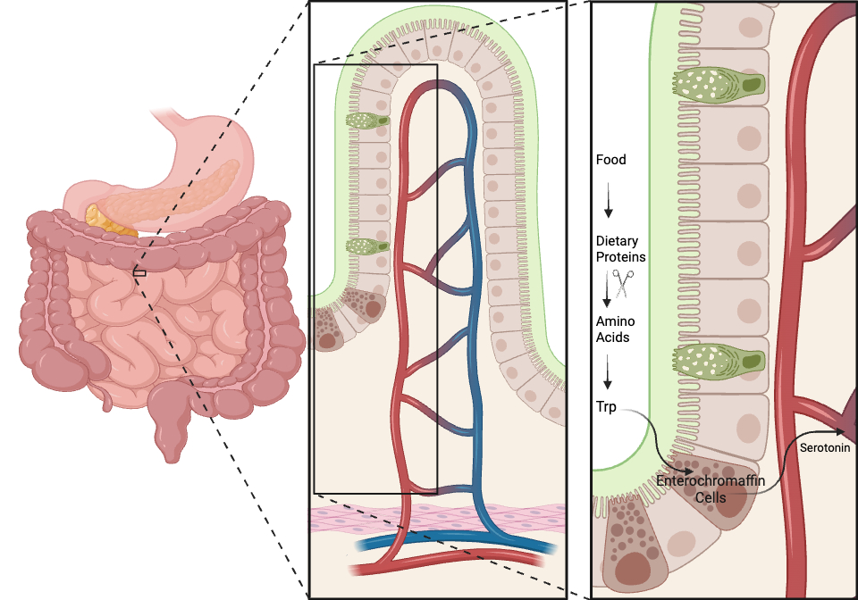
Serotonin is made using a two-step enzymatic process starting from the essential amino acid tryptophan. Tryptophan is obtained from the diet and transported into cells via amino acid transporters. In the gut, enterochromaffin cells convert tryptophan into serotonin through a 2-step enzymatic reaction with 5-hydroxytryptophan (5-HTP) as a metabolic intermediate (each arrow represents an enzyme):
Tryptophan → 5-Hydroxytryptophan (5-HTP)→ Serotonin
The serotonin produced can be stored in enterochromaffin cells or released into the circulation, where it is rapidly taken up by circulating platelets.
Cells other than enterochromaffin cells in the periphery have the enzymatic machinery needed to convert tryptophan into serotonin. However, serotonin production by these cells accounts for less than 1% of peripheral serotonin.
Why Serotonin in the Blood is Soaked Up By Platelets
High levels of free (unbound) serotonin in the blood is dangerous primarily because serotonin is a powerful vasoconstrictor and a potent amplifier of platelet activation and clotting. The body keeps free plasma serotonin extremely low, with only 1% or less of total circulating serotonin not taken up and stored in platelets’ dense granules.
Serotonin Metabolism
Unbound serotonin is eventually metabolized by two different enzymes:
-monoamine oxidase (MAO-A)
-aldehyde dehydrogenase to 5-HIAA (5-hydroxyindoleacetic acid)
The metabolized serotonin is excreted in the urine.
The Roles of Circulating Serotonin
Serotonin acts as a crucial hormone and messenger. The diverse roles of serotonin in the periphery include:
Digestion & Bowel Movements: Serotonin is a key driver of intestinal motility (gut muscle contractions) and fluid secretion, ensuring normal bowel function. The gut also releases extra serotonin to rapidly expel toxic food, which causes nausea and vomiting.
Blood Clotting: Serotonin is stored in blood platelets and released during injury to constrict blood vessels (vasoconstriction), thereby slowing blood flow and initiating the clotting process to stop bleeding.Vagus Nerve Activation: This serotonin binds mainly to 5-HT3 receptors expressed on the terminals of vagal afferent fibers in the gut mucosa. The vagus nerve plays an important role in IACCs. Serotonin activates the vagus nerve to stimulate the nucleus tractus solitarius (NTS) in the brainstem. In mice, this brainstem region was described as an inflammatory thermostat that suppresses inflammatory signals in peripheral tissues.
Immune System: Serotonin is an immunomodulator that regulates immune responses and inflammation, acting as a volume knob that increases and decreases inflammatory responses depending on the:
- Type of immune cells (T cells, Natural Killer cells, macrophages, etc.).
- Which immune cell receptor is activated by serotonin.
- The amount of serotonin present.
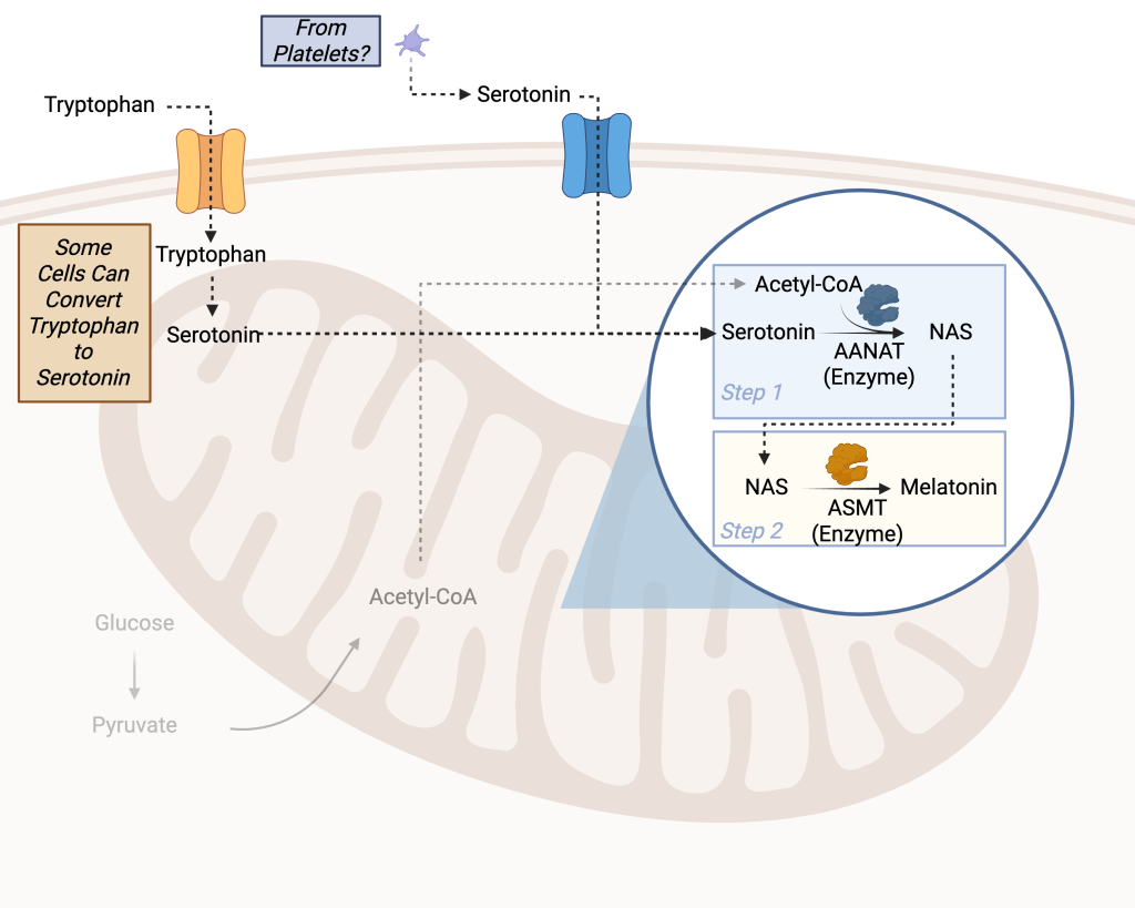
Mitochondrial Melatonin: As we discussed in a previous Substack article, serotonin is used to produce mitochondrial melatonin, a potent master regulator of mitochondrial health and function, which is produced throughout the day and night and therefore does not regulate sleep-wake cycles.
Serotonin Is Used To Make Mitochondrial Melatonin in Peripheral Tissues
Mitochondrial melatonin plays an important role in reducing oxidative stress, one of the most underappreciated cellular pathologies in chronic inflammatory diseases. It can drive endothelial cell dysfunction, stimulate the formation of highly inflammatory immune cells associated with autoantibody production, and impair parasympathetic and sympathetic nervous system function (to name a few).
The predominant source of the oxidative stress-causing free radicals is our mitochondria. Therefore, it makes sense that our mitochondria have many built-in mechanisms that neutralize these free radicals at the source. One of which is the localized production of mitochondrial melatonin.
Mitochondrial Melatonin Production Requires Serotonin Uptake
Almost every cell in your body has the enzymatic machinery needed to make mitochondrial melatonin. However, only a few cell types have the enzymes needed to make the melatonin precursor, serotonin. This makes the production of mitochondrial melatonin partly dependent on the uptake of serotonin from the blood.
Long COVID Reduces Tryptophan and Serotonin Levels in the Blood
One clinical study compared metabolite profiles among Long COVID patients, individuals with acute COVID-19, and those who had fully recovered. They focused specifically on amino acids and their derivatives, which were strongly altered during acute infection.
From this panel, they identified a set of molecules depleted during both the acute and post-acute (Long COVID) phases, with serotonin being the most significantly depleted. However, it was not the only one; the authors also observed that plasma tryptophan levels (the dietary precursor of serotonin) were decreased in patients with Long COVID.
How A SARS-CoV-2 Infection Reduces Tryptophan and Serotonin
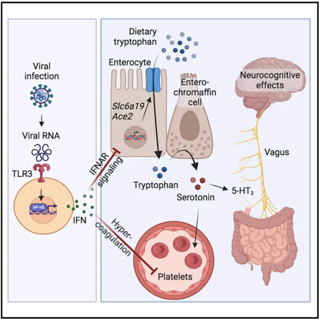
The reduction in tryptophan and serotonin was attributed to the viral infection and the resulting type I interferon-driven inflammation. The sustained inflammation was found to lower serotonin through three specific, interconnected mechanisms:
1. Diminished Intestinal Absorption of Tryptophan: The authors found that viral inflammation reduces the expression of key gut genes (Slc6a19 and Ace2, graphical abstract below) involved in amino acid absorption. This inability to properly absorb dietary tryptophan is reported to cause a shortage of the precursor needed to synthesize serotonin.
2. Impaired Serotonin Storage due to Platelet Depletion: Once synthesized (in the gut), circulating serotonin is normally transported and stored inside blood platelets. However, viral inflammation triggers platelet hyperactivation and hypercoagulability (blood clotting), leading to rapid platelet destruction. With fewer platelets available to store serotonin, circulating levels drop significantly.
3. Increased Serotonin Degradation (Turnover): Any serotonin that remains free-floating in the blood is rapidly broken down by monoamine oxidase (MAO) enzymes. The authors discovered that viral inflammation increases the expression of these MAO enzymes, leading to enhanced turnover and degradation of the remaining serotonin.
The authors also tested an alternative hypothesis: whether tryptophan was being depleted because the body was converting it into a different metabolite called kynurenine.
While kynurenine levels do spike during acute COVID-19, the authors ruled this out as the primary cause for the long-term reduction, noting that kynurenine levels do not stay elevated in Long COVID patients and that blocking kynurenine production in animal models did not restore serotonin levels.
Disclaimer: A letter to the editor was published questioning the accuracy of the results obtained in the manuscript. Thus, we should remain skeptical and wait for other labs to confirm the accuracy of the reported reduction in serotonin.
Reduced Platelet Serotonin in POTS (Postural Orthostatic Tachycardia Syndrome)
A key study found significantly lower serotonin concentrations extracted from platelets in POTS patients, along with clinical symptoms potentially linked to low serotonin. This is because 81% of POTS patients studied show a deficiency in platelet dense granules (where serotonin is stored, among other molecules), resembling a mild δ-storage pool deficiency, leading to lower blood serotonin levels.
This aligns with broader patterns in ME/CFS/Long COVID overlaps, in which inflammation, platelet hyperactivation, or shifts in the kynurenine pathway can reduce serotonin availability. However, it is unknown if low circulating serotonin due to δ-storage pool deficiency is involved in the pathophysiology of POTS.
δ-Storage Pool Deficiency in Long COVID POTS
A study from the same group found that patients who developed POTS following a COVID-19 infection (LCPOTS) have platelet delta granule storage pool deficiency (δ-SPD), presenting with the exact same deficiency as patients who were diagnosed with POTS prior to the pandemic.
However, the authors could not definitively determine whether these Long COVID patients suddenly developed δ-SPD as a direct result of the virus or already had it as an underlying vulnerability.
Serotonin Levels in ME/CFS
A study measured blood metabolites in 70 patients from Italy who experienced chronic fatigue, 45 of whom were diagnosed with ME/CFS. The results revealed that serotonin levels were significantly reduced in the blood compared to healthy controls. However, more robust studies are required to determine the consistency of these results across a larger demographic of ME/CFS patients and whether rescuing circulating serotonin levels can reduce the symptom severity in ME/CFS.
Strategies for Boosting Circulating Serotonin
Medical Disclaimer: This information is for educational purposes only and is not medical advice. Always consult a qualified healthcare professional before starting any supplements, as they can interact with medications, have variable quality and effects, and should be used carefully (with proper dosing, reputable brands, and monitoring).
Based on the aforementioned Long COVID study results, the authors propose several pre-clinical (animal model) treatments to restore serotonin levels and address its downstream neurocognitive effects.
5-HTP Supplementation: Administering 5-hydroxytryptophan (5-HTP), the direct precursor to serotonin, successfully rescued circulating serotonin levels because it completely bypasses the body’s need to absorb dietary tryptophan.
Glycine-Tryptophan Dipeptide Diet: Feeding mice a diet containing a glycine-tryptophan dipeptide enabled tryptophan to be absorbed via alternative dipeptide transporters in the gut, bypassing the primary transporters blocked by viral inflammation. This dietary intervention successfully restored both systemic tryptophan and serotonin levels.
MAO Inhibitors: Treating mice with phenelzine, a pharmacological inhibitor of monoamine oxidase (MAO) enzymes, prevented the rapid degradation and turnover of circulating serotonin, allowing levels to return to normal.The authors conclude that these findings warrant future clinical trials to assess whether precursor supplementation can successfully prevent or treat the neurocognitive manifestations of Long COVID in humans.
Summary
From stimulating the vagus nerve and regulating inflammation to combating oxidative stress and supporting mitochondrial health, serotonin is much more than a neurotransmitter. Despite these important functions, the regulation of peripheral serotonin in infection-associated chronic conditions (IACCs) is understudied. This includes the clinical impact, if any, of restoring circulating serotonin levels on reducing symptoms in those with IACCs.Fluvoxamine, a serotonin reuptake inhibitor (SSRI), was demonstrated to reduce severe fatigue in Long COVID patients. This SSRI is predicted to block serotonin uptake by platelets. Fluvoxamine was chosen over other SSRIs for Long COVID because of its dual-action mechanism.
Beyond blocking the serotonin transporter (SERT) to alter serotonin signaling, fluvoxamine is a potent agonist of the sigma-1 receptor. Activating this receptor confers potent anti-inflammatory effects in the central nervous system, protecting mitochondrial function and reducing the neuroinflammation that drives "brain fog" and physical exhaustion. Thus, the anti-inflammatory properties of fluvoxamine likely outweigh the effects of reducing circulating serotonin levels.
Did you or someone you know have a lab result showing low serotonin levels? Drop a comment below and let us know.


