
The Kynurenine Pathway In Human Health And Disease
Inflammation is a double-edged sword. While it is essential for fighting infections and healing injuries, chronic inflammation in your body can wreak havoc on your brain.
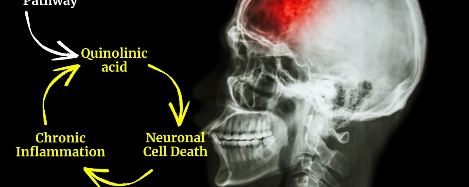
One way it does this is by ‘rewiring’ tryptophan metabolism through the kynurenine pathway, leading to the production of a neurotoxic metabolite called quinolinic acid.
This blog post breaks down everything you need to know about the kynurenine pathway and its role in neuroinflammatory, neurodegenerative, and a subset of neuropsychiatric conditions.
Are you ready to become a kynurenine pathway expert?
Let’s dive in!
The Kynurenine Pathway in CNS Diseases
Peer-reviewed reports suggests that a “dysfunctional” kynurenine pathway occurs in a range of conditions, including ME/CFS (1), Long COVID (2), Parkinson’s disease (3), Alzheimer’s disease (4), traumatic brain injury (5), concussions (5), major depressive disorder (6), bipolar disorder (6), schizophrenia (6), mood disorders (7), and suicidal ideation (8), to name a few.
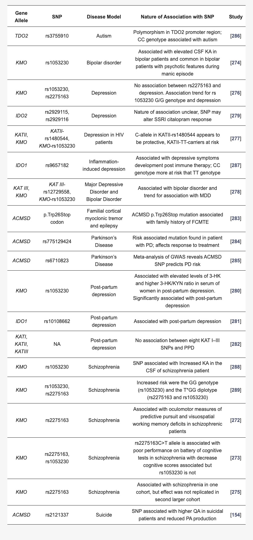
Recent genetic data has identified various DNA mutations in kynurenine pathway genes that might influence the development of various neurodegenerative, neuroinflammatory, and psychiatric disorders (discussed below). However, you could be blessed with perfect mutation-free genes and still suffer from a dysregulated kynurenine pathway. That’s because chronic inflammation can cause the pathway to overproduce a molecule that can be neurotoxic in large amounts.
Let’s zoom in and examine the kynurenine pathway to understand why it plays such a significant role in human diseases.
Pathway Overview
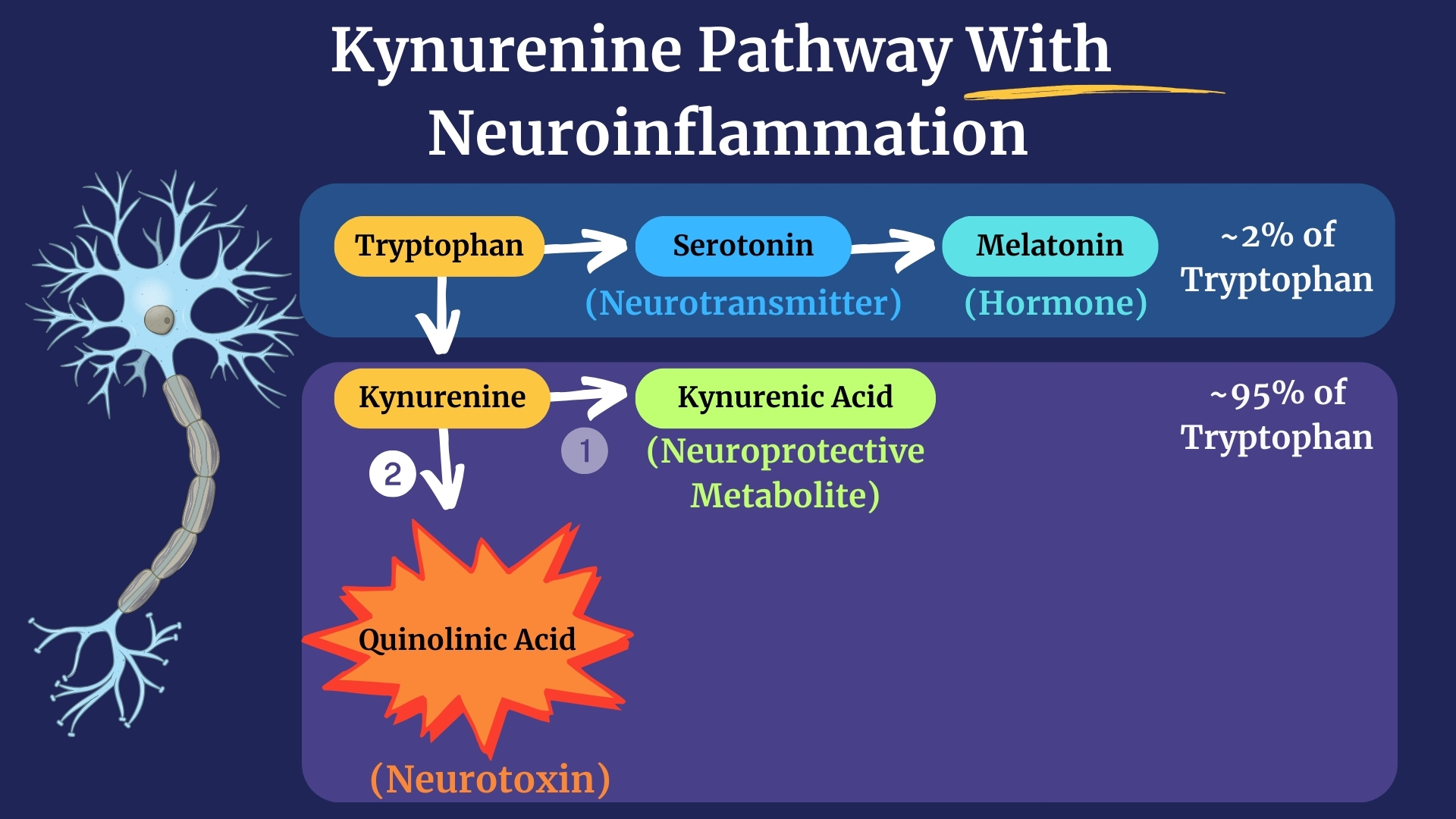
The Kynurenine pathway is a linear metabolic pathway that converts tryptophan, an amino acid found in your diet, into several metabolites, including serotonin, the feel-good neurotransmitter, and melatonin, a powerful antioxidant hormone.
However, under inflammatory conditions, the flow of tryptophan and its downstream metabolites changes, resulting in the accumulation of quinolinic acid, a neurotoxic compound. This disruption in the kynurenine pathway also reduced serotonin and melatonin levels in those with chronic stress, likely contributing to their lack of sleep and anxiety.

Understanding the Pathway, An Analogy
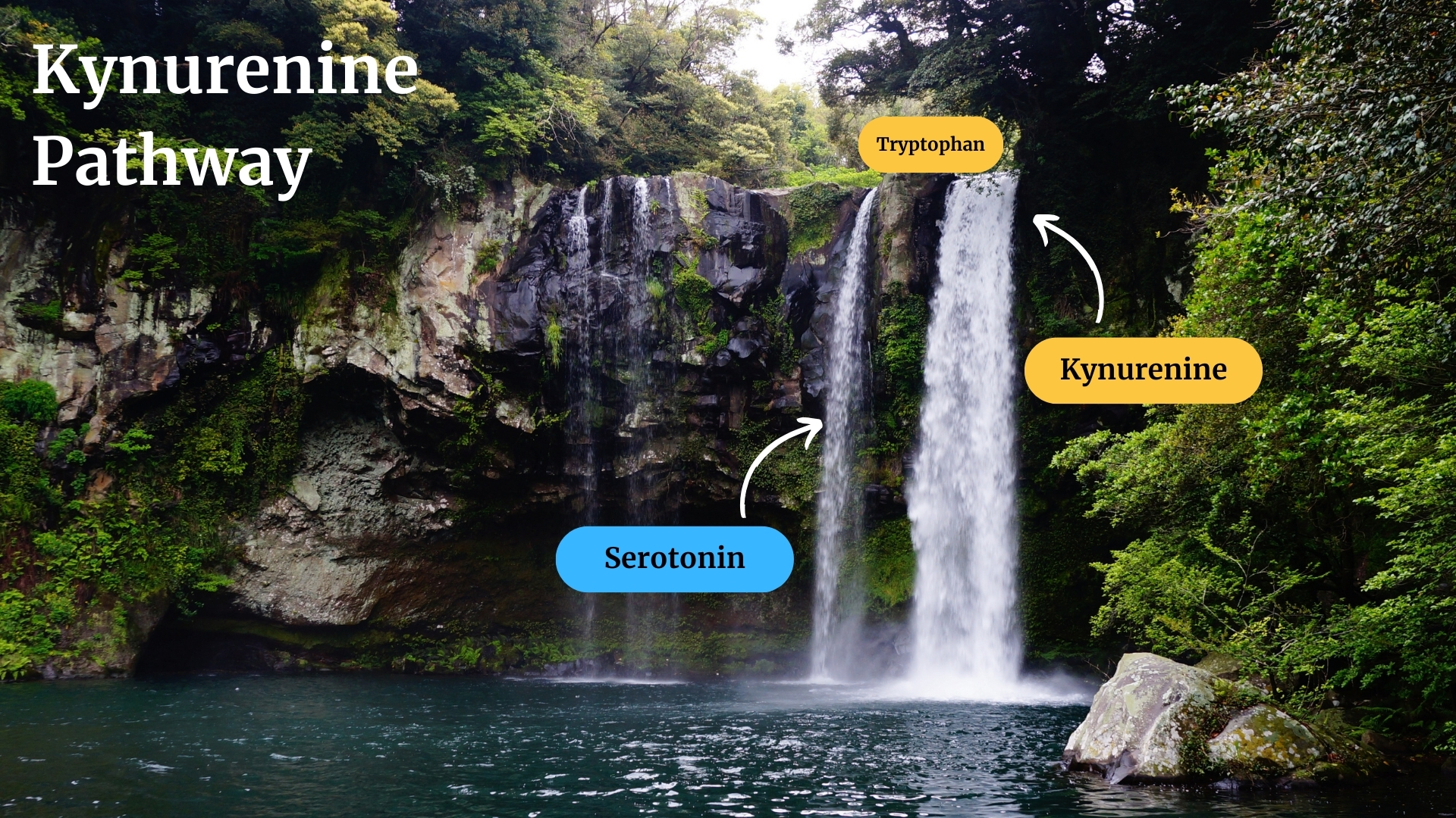
Think of the kynurenine pathway as a waterfall with two main streams (represented below). Tryptophan enters the pathway and rapidly flows downstream, being converted into various metabolites along the way.
Most of the dietary tryptophan is converted to kynurenine, while only 2% of tryptophan is converted into serotonin (9). Kynurenine is a metabolite produced when an enzyme modifies tryptophan.
Let’s take a closer look at the metabolic pathway.
The Kynurenine Pathway Without Inflammation
Under non-inflammatory conditions, the kynurenine branch splits into two directions: one branch produces serotonin and melatonin (light blue shaded box below), while the other produces kynurenine (purple shaded box).
The resulting kynurenine can be converted to kynurenic acid, a neuroprotective metabolite (10). However, inflammation can redirect the flow of kynurenine down a different path.

The Kynurenine Pathway During Chronic Inflammation
Inflammation can reduce the production of kynurenic acid and increase the production of quinolinic acid, a potent neurotoxin (image below) (11). This shift is driven by pro-inflammatory cytokines that modulate the activity of key enzymes in the pathway.
Inflammatory Cytokines and Enzyme Activity
The following inflammatory cytokines increase the activity of indoleamine 2,3-dioxygenase (IDO), the enzyme that converts tryptophan to kynurenine (see image below) (12). The cytokines involved include:
- IL-1β
- IFN-γ
- IFN-α
- TNF-α
- IL-6
- IL-12
While the following cytokines DECREASE (Kynurenine aminotransferase) KAT activity, the enzyme that converts kynurenine to kynurenic acid (13):
- IL-1β
- IFN-γ
- TNF-α

The increased catalytic activity of IDO and the simultaneous decrease in KAT activity redirect the flow of tryptophan through the kynurenine pathway, resulting in the toxic accumulation of quinolinic acid in the CNS.
Subcellular Location of the Kynurenine Pathway
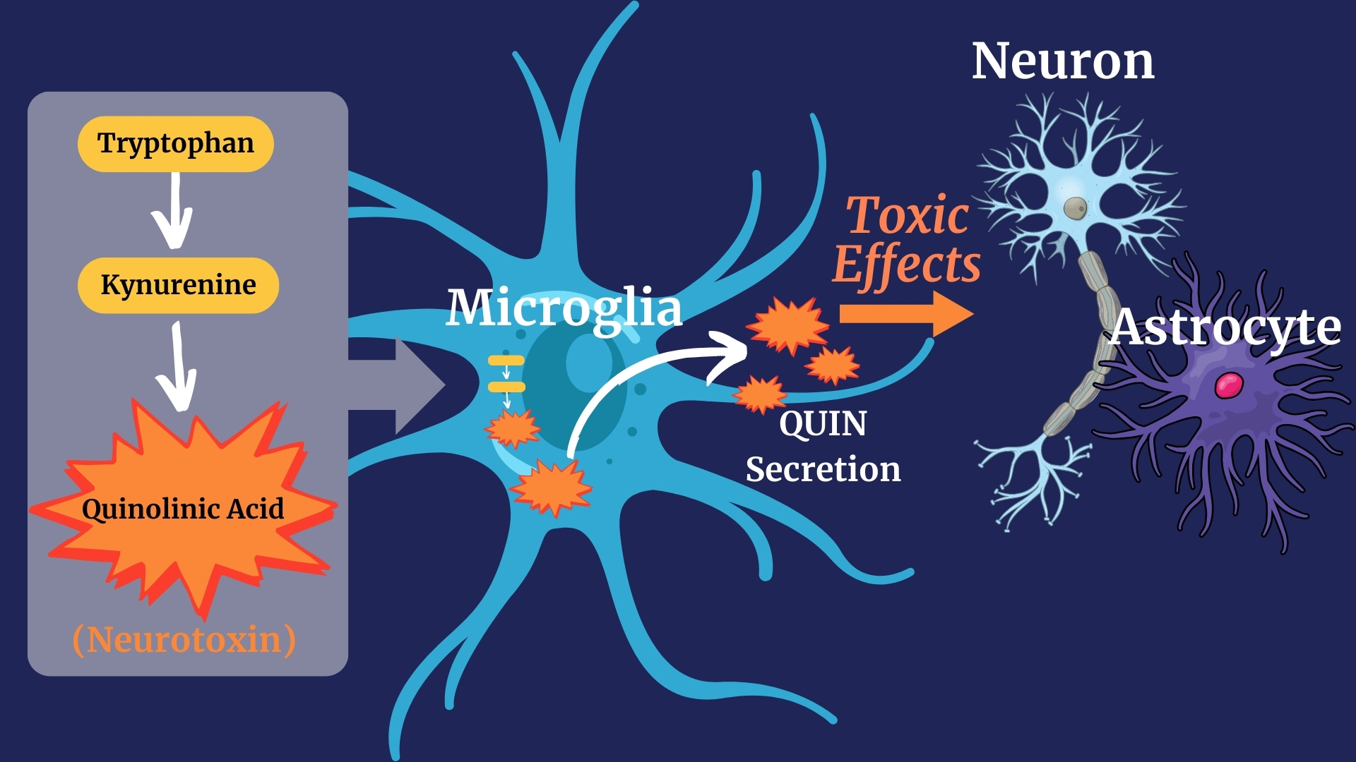
The kynurenine pathway enzymes are located inside the cell (image below) (14). This means the series of biochemical reactions needed to convert tryptophan to kynurenine and other downstream metabolites occurs inside cells.
While the metabolites are produced inside cells, particularly immune cells, they do not necessarily stay inside the cell. Kynurenine, serotonin, melatonin, kynurenic acid, and quinolinic acid can be secreted from cells (released from inside the cell to outside the cell) (15).
Quinolinic Acid Degradation to NAD+ and CO2
There are two intracellular enzymes, not shown in these images, that can diffuse the quinolinic acid bomb:
- aminocarboxymuconate semialdehyde decarboxylase (ACMSD) and
- quinolinate phosphoribosyltransferase (QPRT).
Both enzymes can convert quinolinic acid to harmless byproducts (NAD+, ATP, CO2) (16). Thus, their enzymatic activity can significantly influence the toxic buildup of quinolinic acid inside the cell (16)
However, suppose quinolinic acid is secreted from the cell before it becomes a harmless byproduct. In that case, it will accumulate in the extracellular space (outside the cell), causing toxicity to neurons and astrocytes. This is because no known enzymes outside the cell can break down quinolinic acid (11).
Cell Types Involved in the Kynurenine Pathway
Most cell types can convert tryptophan to kynurenine. However, not all cells can make quinolinic acid. Immune cells such as macrophages, dendritic cells, and microglia (in the CNS) have the enzymes needed to synthesize quinolinic acid during inflammation (17).
While quinolinic acid cannot cross the BBB, tryptophan and kynurenine can. Once inside the CNS, tryptophan and kynurenine can be converted to quinolinic acid by microglia, especially in individuals with neuroinflammation (18).
Astrocytes and neurons lack the enzymatic machinery required to synthesize quinolinic acid (19).
The Consequences of Increased Quinolinic Acid
Quinolinic acid (QUIN) is a potent neurotoxin (20). In 1978, it was observed that injecting QUIN into the brains of mice caused them to convulse (21). Its accumulation was later described to have numerous pathological effects on the CNS, such as:
- Neuronal Excitotoxicity: QUIN stimulates NMDA receptors, causing neuronal excitotoxicity and cell death (20).
- Astrogliosis: QUIN causes astrogliosis, increasing inflammation (22).
- Reactive Oxygen Species (ROS): QUIN increases ROS, leading to oxidative stress (23).
- Lipid Peroxidation: QUIN can damage lipids (24).
- Mitochondrial Dysfunction: QUIN disrupts mitochondrial function (25).
Genetic Factors
Recent genetic testing has identified mutations in genes that regulate the production of kynurenine metabolites involved in depression, schizophrenia, suicidal ideation, Parkinson’s disease, post-partum depression, and more. Below is a table borrowed from this review of the kynurenine pathway.
Kynurenine Pathway-Targeting Drugs
There are at least 14 different kynurenine pathway-targeting drugs used to treat rodent models of Alzheimer’s disease, Huntington’s disease, depression, stroke, brain injury/trauma, epilepsy, Parkinson’s disease, and inflammation-dependent depression (26). However, more work is required to test the efficacy and safety of these drugs in human clinical trials.
The following pharmacological interventions have been found to restore balance to the kynurenine pathway during neuroinflammatory conditions.
| Drug | Drug Type | Condition | Effect |
|---|---|---|---|
|
Escitalopram |
SSRI |
Major Depressive Disorder |
Reduced Quinolinic acid and 3-HK, increased the KA/QA ratio (more KA) (27) |
|
Amitriptyline, Imipramine, Fluoxetine, Citalopram |
SSRI, Tricyclic antidepressants |
Depression |
These antidepressants increase KA abundance in the hippocampus and increases KAT1 and KAT2 expression in the hippocampus (28). |
|
Escitalopram + Celecoxib |
SSRI, COX-2 Inhibitor |
Bipolar Disorder |
Improved response occured with both drugs and peripheral QUIN was reduced (29). |
Adapted from Mithaiwala, M. Santana-Coelho, D. et al. Cells. 2021. PMID: 34205235
Kynurenine Pathway Testing
Measuring the ratio of kynurenine to tryptophan (K/T ratio) in urine, plasma, or CSF is a common method for assessing the catalytic activity of indoleamine 2,3-dioxygenase (IDO), the enzyme that converts tryptophan to kynurenine (30). While this does not directly reflect the amount of quinolinic acid in the CNS, it does provide a proxy for kynurenine pathway activity.
Measuring the ratio of kynurenine to tryptophan requires sophisticated technical expertise and can’t be performed in the clinic. Consult with a healthcare professional to learn if identifying your K/T ratio might be helpful for you.
Conclusion
The kynurenine pathway is a critical metabolic pathway that can be hijacked by inflammation to produce neurotoxic metabolites. This pathway may be among the most important biochemical pathways involved in neurodegeneration, neuroinflammation, and psychiatric conditions. However, there are no medications designed to specifically correct a dysregulated kynurenine pathway.
The accumulation of this potent neurotoxin creates a vicious amplification loop of neuroinflammation, causing neuronal excitotoxicity, astrogliosis, reactive oxygen species production, and mitochondrial dysfunction. These factors can, in turn, cause more neuroinflammation, which leads to more quinolinic acid production. This creates a perpetual cycle of neuroinflammation that can only be stopped by blocking neuroinflammation.

Written By
Matthew Menendez, Ph.D.
Director of Science Communication, Brain Inflammation Collaborative
Dr. Menendez has a passion for empowering patients navigating complex chronic diseases. Through innovative educational strategies, he bridges the gap between cutting-edge research and real-world understanding, helping individuals advocate for better care and access to timely treatments.
Literature Cited
- Kavyani B, Lidbury BA, Schloeffel R, et al. Could the kynurenine pathway be the key missing piece of Myalgic Encephalomyelitis/Chronic Fatigue Syndrome (ME/CFS) complex puzzle? Cell Mol Life Sci. Jul 11 2022;79(8):412. doi:10.1007/s00018-022-04380-5
- Bizjak DA, Stangl M, Börner N, et al. Kynurenine serves as useful biomarker in acute, Long- and Post-COVID-19 diagnostics. Front Immunol. 2022;13:1004545. doi:10.3389/fimmu.2022.1004545
- Venkatesan D, Iyer M, Narayanasamy A, Siva K, Vellingiri B. Kynurenine pathway in Parkinson’s disease-An update. eNeurologicalSci. Dec 2020;21:100270. doi:10.1016/j.ensci.2020.100270
- Liang Y, Xie S, He Y, et al. Kynurenine Pathway Metabolites as Biomarkers in Alzheimer’s Disease. Dis Markers. 2022;2022:9484217. doi:10.1155/2022/9484217
- Dehhaghi M, Heng B, Guillemin GJ. The kynurenine pathway in traumatic brain injuries and concussion. Front Neurol. 2023;14:1210453. doi:10.3389/fneur.2023.1210453
- Marx W, McGuinness AJ, Rocks T, et al. The kynurenine pathway in major depressive disorder, bipolar disorder, and schizophrenia: a meta-analysis of 101 studies. Mol Psychiatry. Aug 2021;26(8):4158-4178. doi:10.1038/s41380-020-00951-9
- Arnone D, Saraykar S, Salem H, Teixeira AL, Dantzer R, Selvaraj S. Role of Kynurenine pathway and its metabolites in mood disorders: A systematic review and meta-analysis of clinical studies. Neurosci Biobehav Rev. Sep 2018;92:477-485. doi:10.1016/j.neubiorev.2018.05.031
- Bryleva EY, Brundin L. Kynurenine pathway metabolites and suicidality. Neuropharmacology. Jan 2017;112(Pt B):324-330. doi:10.1016/j.neuropharm.2016.01.034
- Stone TW, Forrest CM, Darlington LG. Kynurenine pathway inhibition as a therapeutic strategy for neuroprotection. Febs j. Apr 2012;279(8):1386-97. doi:10.1111/j.1742-4658.2012.08487.x
- Stone TW, Darlington LG. The kynurenine pathway as a therapeutic target in cognitive and neurodegenerative disorders. Br J Pharmacol. Jul 2013;169(6):1211-27. doi:10.1111/bph.12230
- Addae JI, Stone TW. Quinolinic Acid and Related Excitotoxins: Mechanisms of Neurotoxicity and Disease Relevance. In: Kostrzewa RM, ed. Handbook of Neurotoxicity. Springer International Publishing; 2022:839-860.
- Xu L, Ling J, Su C, Su YW, Xu Y, Jiang Z. Emerging Roles on Immunological Effect of Indoleamine 2,3-Dioxygenase in Liver Injuries. Front Med (Lausanne). 2021;8:756435. doi:10.3389/fmed.2021.756435
- Hyland NP, Cavanaugh CR, Hornby PJ. Emerging effects of tryptophan pathway metabolites and intestinal microbiota on metabolism and intestinal function. Amino Acids. 2022/01/01 2022;54(1):57-70. doi:10.1007/s00726-022-03123-x
- Guillemin GJ, Cullen KM, Lim CK, et al. Characterization of the kynurenine pathway in human neurons. J Neurosci. Nov 21 2007;27(47):12884-92. doi:10.1523/jneurosci.4101-07.2007
- Badawy AA. Kynurenine Pathway of Tryptophan Metabolism: Regulatory and Functional Aspects. Int J Tryptophan Res. 2017;10:1178646917691938. doi:10.1177/1178646917691938
- Moffett JR, Arun P, Puthillathu N, et al. Quinolinate as a Marker for Kynurenine Metabolite Formation and the Unresolved Question of NAD(+) Synthesis During Inflammation and Infection. Front Immunol. 2020;11:31. doi:10.3389/fimmu.2020.00031
- Schwarcz R, Bruno JP, Muchowski PJ, Wu H-Q. Kynurenines in the mammalian brain: when physiology meets pathology. Nature Reviews Neuroscience. 2012;13(7):465-477.
- Heyes MP, Achim CL, Wiley CA, Major EO, Saito K, Markey SP. Human microglia convert l-tryptophan into the neurotoxin quinolinic acid. Biochem J. Dec 1 1996;320 ( Pt 2)(Pt 2):595-7. doi:10.1042/bj3200595
- Guillemin GJ, Kerr SJ, Smythe GA, et al. Kynurenine pathway metabolism in human astrocytes: a paradox for neuronal protection. Journal of neurochemistry. 2001;78(4):842-853.
- Lugo-Huitrón R, Ugalde Muñiz P, Pineda B, Pedraza-Chaverrí J, Ríos C, Pérez-de la Cruz V. Quinolinic acid: an endogenous neurotoxin with multiple targets. Oxid Med Cell Longev. 2013;2013:104024. doi:10.1155/2013/104024
- Lapin IP. Stimulant and convulsive effects of kynurenines injected into brain ventricles in mice. J Neural Transm. 1978;42(1):37-43. doi:10.1007/bf01262727
- Ting KK, Brew BJ, Guillemin GJ. Effect of quinolinic acid on human astrocytes morphology and functions: implications in Alzheimer’s disease. J Neuroinflammation. Dec 10 2009;6:36. doi:10.1186/1742-2094-6-36
- Iwahashi H, Kawamori H, Fukushima K. Quinolinic acid, alpha-picolinic acid, fusaric acid, and 2,6-pyridinedicarboxylic acid enhance the Fenton reaction in phosphate buffer. Chem Biol Interact. Apr 15 1999;118(3):201-15. doi:10.1016/s0009-2797(99)00080-0
- Stípek S, Stastný F, Pláteník J, Crkovská J, Zima T. The effect of quinolinate on rat brain lipid peroxidation is dependent on iron. Neurochem Int. Feb 1997;30(2):233-7.
- Bordelon YM, Chesselet MF, Nelson D, Welsh F, Erecińska M. Energetic dysfunction in quinolinic acid-lesioned rat striatum. J Neurochem. Oct 1997;69(4):1629-39. doi:10.1046/j.1471-4159.1997.69041629.x
- Mithaiwala MN, Santana-Coelho D, Porter GA, O’Connor JC. Neuroinflammation and the Kynurenine Pathway in CNS Disease: Molecular Mechanisms and Therapeutic Implications. Cells. Jun 19 2021;10(6)doi:10.3390/cells10061548
- Halaris A, Myint AM, Savant V, et al. Does escitalopram reduce neurotoxicity in major depression? J Psychiatr Res. Jul-Aug 2015;66-67:118-26. doi:10.1016/j.jpsychires.2015.04.026
- Kocki T, Urbańska EM, Kocki J, et al. ProLonged therapy with antidepressants increases hippocampal level of kynurenic acid and expression of Kat1 and Kat2 genes. Pharmacol Rep. Aug 2018;70(4):737-745. doi:10.1016/j.pharep.2018.01.004
- Castillo MFR, Murata S, Schwarz M, et al. Celecoxib augmentation of escitalopram in treatment-resistant bipolar depression and the effects on Quinolinic Acid. Neurology, Psychiatry and Brain Research. 2019;32:22-29. doi:10.1016/j.npbr.2019.03.005
- Lionetto L, Ulivieri M, Capi M, et al. Increased kynurenine-to-tryptophan ratio in the serum of patients infected with SARS-CoV2: An observational cohort study. Biochimica et Biophysica Acta (BBA) – Molecular Basis of Disease. 2021/03/01/ 2021;1867(3):166042. doi:https://doi.org/10.1016/j.bbadis.2020.166042

